- Get link
- X
- Other Apps
As part of medical research, scientists often resort to a modeling method that artificially creates the conditions and environment in which viruses and other pathogens are added, from which experts then try to develop medicines. The research laboratory at the University of Drekstel, specializing in bioprocessing, most recently, thanks to the 3D printing method, has created a model of tumor tissue that more closely imitates this material than the more traditional two-dimensional artificially created cultures.
The main objective of this study was to create an environment that would allow a better understanding of the growth process of the blast and, more importantly, the search for ways to effectively treat them.
Tumor tissues grown in the laboratory can vary in size, the total area of the lesion, as well as the shape of their cells and cell composition. It often happens that scientists seemingly find a potential treatment for cancer and successfully apply it on artificially grown tumors whose artificial cells do not resist the drug. But in real conditions all this leads only to unfulfilled expectations and hopes.
Dr. Wei San’s research at Drextel’s University was based on analyzing the most appropriate method for printing HeLa cells (a known strain partially responsible for developing cervical cancer) and searching for the necessary supported matrix of proteins that can usually be found in the body near the affected area. After that, the team of scientists compared how 3D-tumor cells were chemically resistive compared to a 2D culture consisting of the same cells.
Scientists note that heat and complex mechanical effects are important components of the production of living 3D cells, so for the initial tests, the researchers chose a process of linear cell printing, but in the future they are going to improve and change this process for more efficient use.
The selection of the desired temperature is a key parameter for the correct viscosity of fibrin, alginate and a special gelatin mixture (base), which was chosen as a protein-imitating material and in which the cells themselves usually grow. If the temperature is higher than necessary, then in the process of printing the cells of the cancer will die. If the temperature is too low, more force will be needed to compress the gelatin base. Again, the cells will die. The search for suitable solutions to these questions led scientists to more concentrated attention on the printing process itself.
Printing cells allows scientists to mimic a model of natural growth. The tubules in the printed structure are used to deliver oxygen, nutrients, and waste products in the same way as real tissues. After eight days of growth, scientists noted that 90 percent of the cells remained alive, and the tissue itself formed a spheroidal structure of cells with strong cell-cell connections. In the case of a 2D environment, the cells would remain in their original flat position. In addition, it was observed that three-dimensional printed cells show a higher level of growth, as is usually the case with real tumor cells.
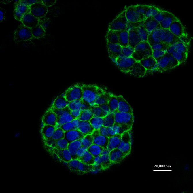
The tumor spheroids marked with fluorescent paint after 8 days from the moment of their printing. Cells are marked blue, green is extracellular material.
After it was proved that this process allows you to create high-quality living tissue, the next step for scientists was even more important: checking the resulting tissue for adequacy of the response of medical agents against cancer. In tests using paclitaxel, tumor spheroids were more chemically resistant than two-dimensional tissue. Such a result may indicate the accuracy and adequacy of the response of 3D-cells to a medical drug. The same result can be noted with real live tumors.
The researchers' future plans include printing fabrics consisting of various types of cells, as well as the imposition of some printed cells on other printed tissues in order to simulate the process of their real growth.
It should be noted that Dr. San created his own 3D printer back in 2002. Since then, he and his team from the bio-production lab can create tissue and bone samples. The results of the work done by scientists recently shared in the pages of the journal Biofabrication.
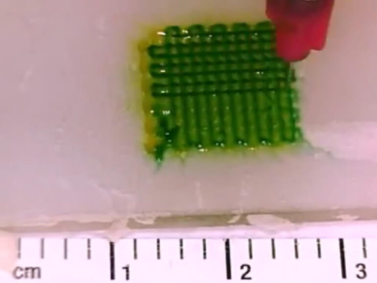
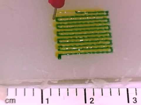
In the video below you can see the printing process of multilayer biological structures.
The article is based on materials .
Comments
Post a Comment